CT Imaging
Instruments
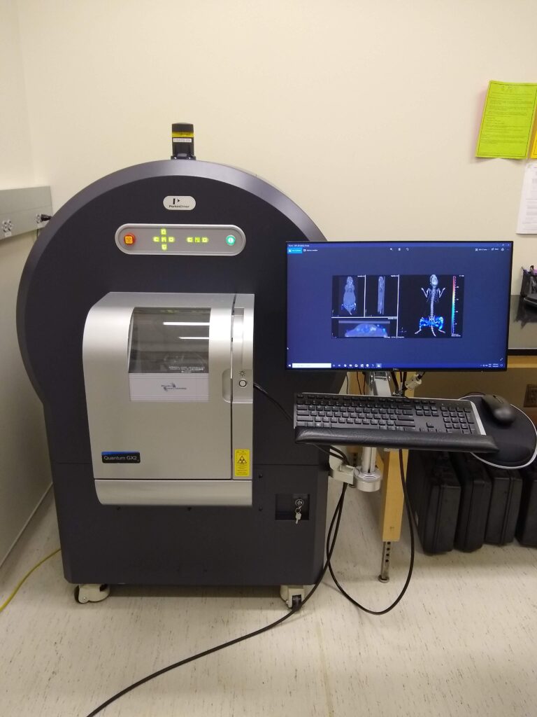
| PerkinElmer Quantum GX2
The Quantum GX2 microCT scanner is a true multispecies preclinical imaging system, offering the flexibility to enable longitudinal in vivo imaging as well as ex vivo sample scanning. Main Imaging Features
|
 |
| GE eXplore CT120
High-performance, high-throughput small animal In Vivo MicroCT scanner designed for high-quality scanning for a wide variety of applications. It is designed to help visualize, quantify, and characterize anatomical parameters in small animals such as mice and rats. Main Imaging Features
|
 |
| Scanco uCT40 Specimen CT
The SCANCO µCT 40 scanner is a high resolution desktop cone-bean X-ray scanner designed for specimens with a top resolution of of 6 µm. In addition to the high resolution capabilities, it also offers a larger specimen size (36 mm diameter, 8 cm specimen length).
|
 |
Imaging Applications and Examples
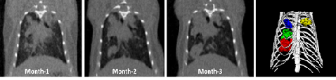
| In-Vivo Gated Lung CT
|
In-vivo Gated Lung CT
|
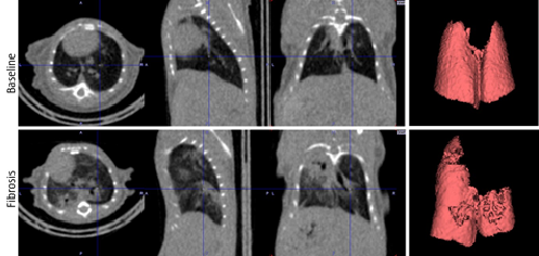
| Ex-Vivo High Resolution Lung CT
|
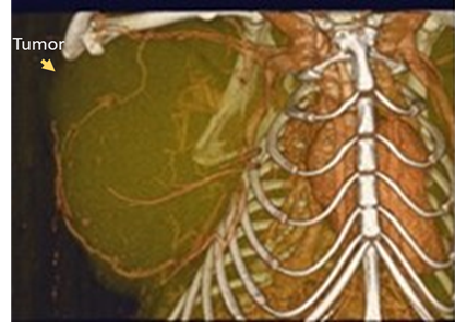
In-Vivo Contrast Enhanced CT
|
| High Resolution Specimen CT
|
|


Other Sample studies
- Bone regrowth after implant
- Multiple dental applications (microfractures, implants, crown effectiveness, anatomy, etc.)
- Arthritis models
- Contrast enhanced soft tissue scans (i.e. embryo, cartilage, muscle, etc.)
- Calcification in human cardiac arteries
- Bone density loss in calvaria
- Vascular and alveolar imaging
- Materials testing
Study Initiation, Training and Scheduling
Training is not available on these systems. To initate a CT imaging study, please send an email to bricsai@med.unc.edu to schedule a meeting and register your project by following the study initiation link. You can also see our study initiation page for more details.