Tarran Lab Images
 |
Figure 1. Role of the A2b signaling pathway in ASL hydration. ATP is secreted from cells basally or following shear stress and is hydrolyzed by ecto-enzymes to AMP. The 5’ ecto-nucleotidase (5’NT; 1) then degrades AMP to ADO, which activates A2b receptors (A2b-R, 2) that are coupled to G-proteins (Gs) and adenylyl cyclase (AC) to raise local concentrations of intracellular cAMP, resulting in activation of CFTR and inactivation of ENaC. ADO is then degraded to inosine by adenosine deaminase (ADA, 3) and taken up into the cell by nucleotide transporters (eNT1, 4). |
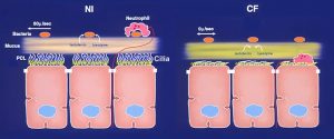 |
Figure 2. Schematic model of early pathogenic events in CF airways. Left, on normal airway epithelia, a thin mucus layer resides atop the periciliary liquid layer (PCL), which is maintained by active ion transport (e.g. Na+ & Cl- channels). The presence of the low viscosity PCL facilitates efficient mucociliary clearance and allows free movement of neutrophils which engulf inhaled bacteria. Right, excessive CF volume depletion caused by abnormal ion transport removes the PCL, mucus becomes adherent to epithelial surfaces, and mucus transport slows/stops. Due to the concentration of mucus, neutrophil movement also becomes impaired and natural antibactierial agents such as lactoferrin and lysozyme are insufficient to kill bacteria.
|
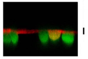 |
| Figure 3. RSV infections inhibit PCL homeostasis in CF airways. XZ confocal image of PCL (red) covering RSV-gfp-infected CF ciliated cells (green). This culture has lost the ability to regulate PCL volume due to RSV-induced upregulation of ecto-ATPases, which deplete the ASL of ATP, a vital signaling molecule in CF ASL. |
 |
| Figure 4. Simultaneous imaging of ERα and Fura-2, as an indicator of intracellular Ca2+ in BHK cells. Images of Fura-2-loaded BHK cells (green) and BHK cells expressing ERα linked to orange fluorescent protein (mOr; green/orange). |
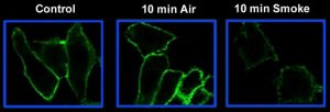 |
| Figure 5. Cigarette smoke induces removal of CFTR from the plasma membrane. BHK cells constitutively expressing CFTR are labeled with a green antibody against an extracellular portion of CFTR. After cigarette smoke exposure, the amount of CFTR in the plasma membrane is markedly diminished. |
| Movie 1. The structure of short palate lung and nasal clone 1 (SPLUNC1). |