Ever wanted to see what a neuron looks like? Can you visualize the vessels that drain excess fluid from a heart?
Images by UNC Lineberger Comprehensive Cancer Center researchers were among those chosen for an exhibition of work by UNC School of Medicine scientists at the North Carolina Museum of Art.
“The Art of Science and Innovation,” exhibition was designed to reveal the “hidden beauty” of biomedical research. It will be on display through Monday, Jan. 14, in the museum’s East building.
“We use art to understand science,” said UNC Lineberger’s Kathleen Caron, PhD, professor and chair of the UNC School of Medicine Department of Cell Biology & Physiology. “(The images in this exhibit) represent the laboratories where images and seeing are at the heart … of how (these scientists) answer questions,” she added.
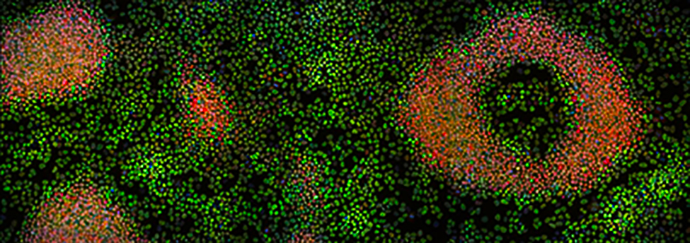
Caron explained the historical connection between art and science, as well as current basic and translational research efforts that are ongoing at the UNC School of Medicine during a tour of the exhibition. The exhibition includes images of neurons in a model of the brain, genetic material spilling out of the E. coli bacterium, and of the genetic material of a cancer-linked virus binding the DNA of its host.
“I would totally put this in my living room,” Caron said, pointing to one vibrantly colored image that depicted cilia, hair-like structures that help sweep mucus out of the airway. The piece, “Lung surface; protected by a mucus blanket,” was created by Mehmet Kesimer, PhD, and Sarah Keilson, medical illustrator. When this system does not function properly, people can develop diseases like cystic fibrosis.
“You know when you get really sick? Here’s the culprit,” Caron said, pointing to an image of genetic material spilling out of E. coli. UNC Lineberger’s Jack Griffith, PhD, professor in the UNC School of Medicine Department of Microbiology and Immunology, used electron microscopy to capture the image. The tool allows for visualization of molecular properties, down to the coiled DNA from a single bacterium.
“This is the difference between light and electron microscopy – we see at a very minute level,” she said.
“One of the missions of the North Carolina Museum of Art is to connect art, nature and people, and this is a fantastic example of connecting nature and art with the researchers who have made it possible,” said Kat Harding, public relations manager for the museum.
To view more images click here
News courtesy of Lineberger