Optical Imaging
Instruments
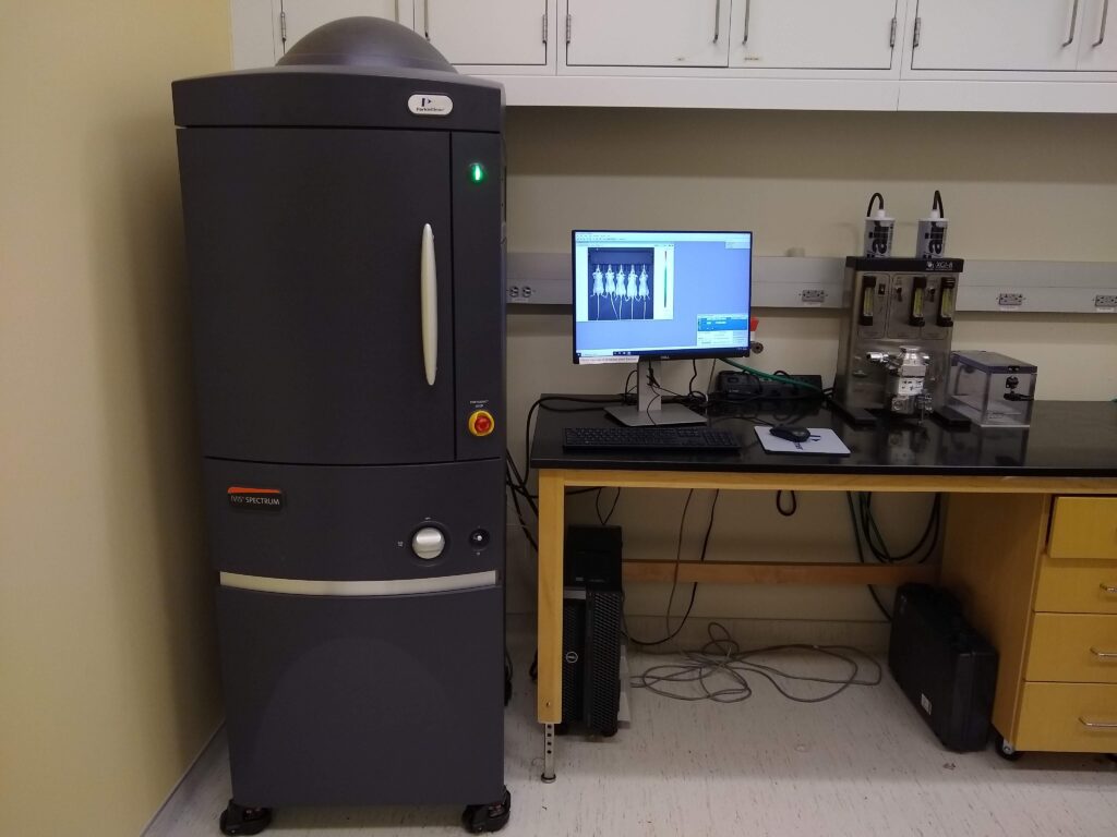
| IVIS Spectrum
Located in Marsico Hall, the IVIS® Spectrum (PerkinElmer Inc.) in vivo imaging system combines 2D optical and 3D optical tomography in one platform. This instrument can be used in conjunction with our Quantum GX2 CT system to combine 3D optical with CT imaging Main Imaging Features
|
 |
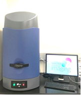
| IVIS Lumina
The IVIS-Lumina (PerkinElmer Inc.) provides highly sensitive bioluminescence and fluorescence imaging with physiological relevant molecular and functional information. The IVIS Lumina is located in the Genetic Medicine Building room UB61 and facilitates optical imaging studies with animals located only in the GMB vivarium. Main Imaging Features
|
|
| AMI HT
The Ami system is capable of providing both bioluminescence and fluorescence imaging on animals and tissue specimens for preclinical research. The system is housed in Marsico Hall SAI imaging facility. The Ami system is very similar to the IVIS system in terms of imaging performance and operation. Main Imaging Features
|
|

Imaging Applications and Examples

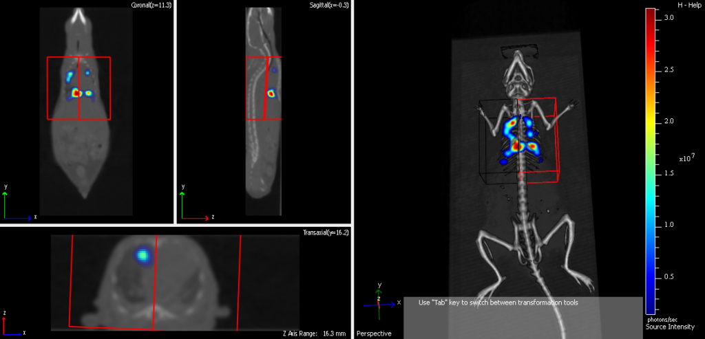
3D bioluminescence imaging of brain tumor pre-surgery (PIs: Kristy Ainslie and Shawn Hingtgen, courtesy of Liz Gurysh)
3D fluorescence imaging and volumetric analysis of primary and metastatic mouse lung tumors (PI: Chad Pecot, courtesy of Emily Harrison)
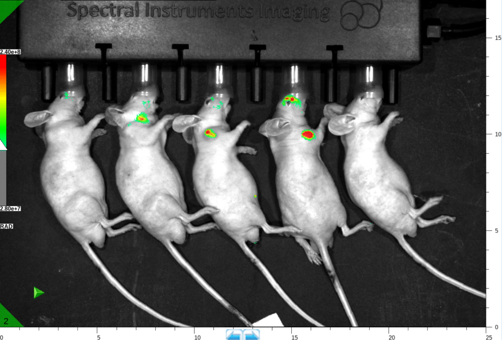
2D fluorescence hypoxia optical imaging of FaDu tumor following four hours of carbogen breathing (PI: Hong Yuan)

Fluorescence imaging of knee inflammation post-surgery (PI: Lara Longobardi, courtesy of Huseyin Ozkan)
Other Optical Imaging Study Examples
- Infectious Disease
- Metabolic Disease
- Neurology
- Gene Therapy
- Stem cell Biology
- Cardiovascular Disease
- Drug metabolism
- Cherenkov radiation imaging
Study Initiation, Training and Scheduling
The SAI offers monthly group training on all optical imaging systems and private training can be arranged (will include fee for machine time and staff support. Please click on this link to learn more. To initate a new optical imaging study, please send an email to bricsai@med.unc.edu to schedule a meeting and register your project by following the study initiation link. Please contact us at bricsai@med.unc.edu or call us at 919-966-2855 with any questions about your optical study. Optical imaging can be scheduled after training by SAI staff through iLab. Please follow the instructions at the Schedule a Study link for more details or contact us at bricsai@med.unc.edu with questions.
Cost
The current internal rate for optical imaging is $48/hr. Imaging time is highly dependent upon the study protocol so please contact the SAI for budget information. You can also see our rates at this link.