Previous SAI Seminars
2014
Date: Tuesday, Feb 4th, 2014
Time: 1:00-2:00 PM
Seminar Title: Contrast and Conquer: Gold and X-Rays from Vascular Visualization to Curing Cancer
Speaker: Richard Powell, Ph.D., Research Director, Nanoprobes, Inc.
Location: BRIC conference, (Medical Research D Building, BRIC Facility building)
Date: Tuesday, October, 14th, 2014
Time: 1:00-2:00 PM
Seminar Title: Developing the right PET agents for targeted imaging
Speaker: Zibo Li, Ph.D, Director, Radiochemistry and Cyclotron Facility at BRIC, UNC
Location: Marsico Hall, Room 2004 (conference room)
Date: Tuesday, December 9th, 2014
Time: 1:00-2:00 PM
Seminar Title: New Approaches for Magnetic Resonance Imaging and Spectroscopy
Speaker: Tamara Branca, Ph.D, Assistant Professor, Department of Physics and Astronomy, BRIC, UNC
Location: Marsico Hall, Room 2004 (conference room)
2015
Seminar Title: Contrast enhanced ultrasound imaging and its role in preclinical biomedical research
Speaker: Paul Dayton, Ph.D, Professor, Department of Biomedical Engineering, UNC
Date: Thursday, March 12th, 2015
Time: 1:00-2:00 PM
Location: Marsico Hall, Room 6004 (conference room)
Seminar Title: New Development on microCT Imaging
Speaker: Rasesh Kapadia, President, Scanco USA, Inc.
Date: Wednesday, April 15th, 2015
Location: Marsico Hall, Room 3004 (conference room)
Seminar Title: Developing Stem Cell Drug Carriers for Cancer Therapy
Speaker: Shawn Hingtgen, Ph.D., Assistant Professor, School of Pharmacy, Biomedical Research Imaging Center
Date: Tuesday, May 19th, 2015
Time: 1:00-2:00 PM
Location: Marsico Hall, Room 2004 (conference room)
Dr. Hingtgen’ s lab focuses on developing stem cell-based therapeutic methodology for cancer treatments. Their group has utilized extensively non-invasive imaging methods in stem cell research, particularly, optical imaging including both bioluminescence and fluorescence imaging. Dr. Hingtgen will talk to us his research on developing stem cell drug carriers, and how optical imaging has been incorporated in the various stages of the study as an important research tool.
2016
Seminar Title: Small Animal Imaging with the LI-COR Pearl Trilogy: Sensitive Detection of Near-IR Fluorescence and Bioluminescence
Speaker: Janeen L. Vanhooke, Ph.D., Li-Cor Biosciences
Date: February 23rd, Tuesday, 2016
Time: 1:00-2:00 PM
Location: Marsico Hall, Room 4004 (conference room)
Abstract: Janeen Vanhooke from Li-Cor will give a talk mainly focusing on near-infrared fluorescence imaging, which provides much deeper tissue penetration and low auto-fluorescence artifacts.
Date: Tuesday, March 15th, 2016
Seminar Title: Preclinical imaging with new cryogen-free superconducting MRI: Multi-modality opportunities and challenges
Speaker: Gilberto Prudencio, President, MR Solutions Americas
Time: 1:00-2:00 PM
Location: Marsico Hall, Room 3004 (conference room)
Date: Tuesday, March 22th, 2016
Seminar Title: High-end Solutions for Quantitative Translational PET Imaging: PET/CT & PET/MRI
Speaker: Illes Muller, Mediso USA
Time: 1:00-2:00 PM
Location: Marsico Hall, Room 3004 (conference room)
Date: Tuesday, May 17th, 2016
Seminar Title: In vivo multi-modality imaging of disease and therapy: Optical, microCT, and PET imaging
Speaker: Vivek R. Shinde Patil, PhD, Senior Manager, Preclinical Technical Applications
Time: 1:00-2:00 PM
Location: Marsico Hall, Room 4004 (conference room)
Abstarct: The seminar will be mainly focusing on optical imaging. Dr. Patil from PerkinElmer will give us updates on the optical imaging and other preclinical imaging techniques. As many of you have been using the IVIS optical imaging systems in our facility, it will be interesting to find out what are the new technologies and multimodality imaging options available now to researchers. In addition, we will be hearing how these technologies can be efficiently applied to various research fields, including oncology, infectious disease, neurobiology, stem cell and transplantation research, cardiovascular disease, gene therapy, etc. Dr Patil has years of experiences on preclinical optical imaging and imaging probes, if you have any questions on using the IVIS optical system, this will be a good chance to talk to him.
Date & Time: Tuesday, August 16th, 2016, 1-2pm
Location: Marsico Hall, room 2004
Title: Image-guided Radiation on Small Animals: Getting the Most Relevant Results on Preclinical Models
Speaker: Adrian Treverton, COO of Xstrahl, LTD
Seminar abstract: Effective translation of novel treatment methods requires rigorous pre-clinical studies and evaluation, however, until recently, the nonexistence of small, rodent sized modern radiation therapy equipment has presented a serious impediment to the progress of radiation oncology research. Many pre-clinical studies simply have not been conducted because the necessary technology, such as conformal irradiation and image guidance, did not exist to provide laboratory animals irradiation conditions that parallel those of the modern clinic. The seminar will be exploring how image guided small animal radiation with the Small Animal Radiation Research Platform (SARRP)- can transform pre-clinical radiation research.
Date & Time: Tuesday, November 1st, 2016, 1-2pm
Location: Marsico Hall, room 5004
Title: Explore the Possibilities With Magnetic Particle Imaging
Speaker: Dr. Patrick Goodwill, CTO of Magnetic Insight
Join us for a presentation on the newest molecular imaging technology since PET. The team from Magnetic Insight will present on the basics of MPI and application data providing unique solutions in:
- Cell Tracking Models
- Functional Tumor Imaging
- Localized Hyperthermic & Theranostic Imaging
- Functional Vascular Imaging
Unlike Magnetic Resonance Imaging (MRI), Magnetic Particle Imaging (MPI) is a unique, ultra-sensitive, high resolution molecular imaging approach that measures the magnetic fields generated by iron oxide labeled nanoparticles to form tomographic images. MPI harnesses the flexibility of iron oxide nanoparticles to label cells, as targeted probes, or freely flowing through the vasculature.
(Coffee and snacks will be served)

Date: Tuesday, December 13th, 2016
Time: 1:00-2:00 pm
Location: Marsico Hall, room 2004
Title: Multispectral Optoacoustic Tomography (MSOT) In Vivo Imaging: Principles and Applications
Speaker: Clinton Hupple, Application Scientist, iTheraMedical
Multispectral Optoacoustic Tomography (MSOT) is a powerful new molecular imaging technology which combines high-resolution real-time ultrasound detection with the versatile specificity of optical contrast, thus giving information about physiological processes in vivo at a molecular and cellular level. Uniquely, it provides the capacity to detect either endogenous signals of interest such as oxy-/deoxy-hemoglobin or tissue contrast from exogenously administered agents including nanoparticles and fluorescent dyes or proteins. This presentation will focus on technical development and imaging applications in the following area:
- Oncology applications
- Particle biodistribution applications
- Whole body imaging
- High resolution raster scanning optoacoustic mesoscopy (RSOM) imaging
Date & Time: Tuesday, September 17th, 2019, 1-2pm
Location: Marsico Hall, Room 2004
Title: Cryo-Fluorescence Tomography, A Technique to Bridge Multi-Resolution Multi-Modal Assays
Speaker: Mohammed Farhoud, EMIT Imaging, Inc.
Abstract:
Many human cancer cell lines have been engineered to express fluorescence so that in vivo imaging can be used to monitor and stage disease progression. Following in vivo imaging, traditional histo-pathology can be performed to validate in vivo measurements. However, a gap in sensitivity and resolution between in vivo and ex vivo techniques may make it hard to characterize an animal model. Traditional ex vivo techniques only focus on small sample sizes while allowing for high-resolution evaluation and characterization. Using a Cryo-Fluorescence Tomography (CFT) imaging approach, an imaging modality based on serial slicing and off-the-block fluorescence imaging, we can now bridge the gap between in vivo and ex vivo resolution of the entire animal. This new technique and imaging procedures will be described with various research examples.
———————–
(Snacks and coffee will be served)

2021
NEW Imaging Technology – In-vivo Fluorescence Endomicroscopy
Date: June 9th, 2021, Wednesday
Time: 2:00 -3:00 PM ET
Title: In-vivo Fluorescence Endomicroscopy
Speaker: Mohammedayaz Rangrez, PhD, Applicant Manager, OptiScan North America
Zoom Link:
Obtain a meeting link by emailing us at bricsai@med.unc.edu
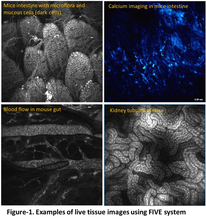
Abstract: Histology is one of the most important tools in the armaments of a biologist. It can uncover the cellular mechanisms behind a disease, explain toxicity of a substance or show the effectiveness of a treatment. However, the method has not changed significantly in the last 250 years. Though standardized, this process is highly invasive and may induce severe artifacts. Fluorescence in vivo endomicroscopy (FIVE) is a virtual histology device that can capture real time, histology equivalent images, directly from a live animal, without sacrificing the tissue (Figure 1). FIVE is also referred to as “virtual biopsy” as one can look at living tissue architecture without performing physical biopsy. Additionally, this device can access organs and tissues in larger animals that do not fit on 2-photon microscope stage. This live zoom meeting will include theoretical discussion and practical demonstration of the FIVE-2 machine and its research applications. If you are working with animals, it may have great learning value for your research. Use the link above to join this zoom meeting.