James Bear, PhD and Klaus Hahn, PhD, Directors
The UNC-Olympus Research Imaging Center is a unique academic-commercial partnership that brings together state-of-the-art biology, chemistry and engineering from UNC-Chapel Hill together with world-class microscopes and imaging from Olympus. Together, these partners are unraveling biological problems that are critical for improving human health and deepen our understanding of the natural world.
In the UNC-Olympus Research Imaging Center, a group of principal investigators drawn from diverse departments across the university collaborate on joint projects that cross the traditional boundaries of discipline and method. These cross-disciplinary approaches are facilitated by access to leading-edge microscopes, and direct collaboration with Olympus personnel on imaging experiments using prototype microscope designs and established commercial instruments. The Center provides a crucial real world testing ground for new Olympus products, and ultimately the creation of the next generation of microscopes.
The Olympus Center is an imaging research ‘incubator’ where investigators develop collaborative, cross-disciplinary projects that would not be possible without the expertise of multiple labs and the imaging resources of the Center. In addition to collaborative science, the Center also serves as a vehicle for educational activities such as workshops in specific imaging techniques.
UNC-Olympus Research Imaging Center Principal Investigators
Nancy Allbritton, Distinguished Professor Chemistry, and Chair of Biomedical Engineering
Vicki Bautch, Professor/Director for Developmental Biology, Biology
Jim Bear, Associate Professor, Cell Biology and Physiology
Richard Cheney, Professor, Cell Biology and Physiology
Tim Elston, Professor, Pharmacology
Shawn Gomez, Associate Professor, Biomedical Engineering
Stephanie Gupton, Assistant Professor, Cell Biology and Physiology
Klaus Hahn, Distinguished Professor, Pharmacology
Shawn Hingtgen, Assistant Professor, Eshelman School of Pharmacy
Ken Jacobson, Professor, Cell Biology and Physiology
Gary Johnson, Professor and Chair, Pharmacology
John Rawls, Associate Professor, Cell Biology and Physiology
Jon Serody, Distinguished Professor, Medicine
Steven Soper, Professor, Biomedical Engineering/Chemistry
Rich Superfine, Professor, Physics and Astronomy
Anne Taylor, Assistant Professor, Biomedical Engineering
Images by some of the Principal Investigators involved in the research at the UNC-Olympus Research Imaging Center
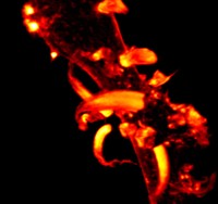
This image shows actin-rich fungipod protrusions forming on a human white blood cell after stimulation with yeast cells. Fungipods are novel protrusive structures discovered by Aaron Neumann in Ken Jacobson’s lab that may participate in clearing fungal infections such as those observed in patients with compromised immune systems.
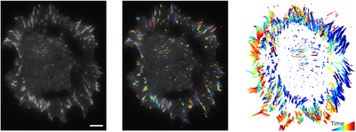
This image above illustrates a novel method developed by Shawn Gomez’s lab (in collaboration with Klaus Hahn’s group) of quantifying the dynamics of cell adhesion structures over long time periods. These structures, called focal adhesions, are regulated by signaling events that become disrupted or misregulated in metastatic cancer and other disease states. The left panel shows the starting image, the middle panel shows the results of a computer-vision based image segmentation and the right panel shows those adhesions over time (coded in color).
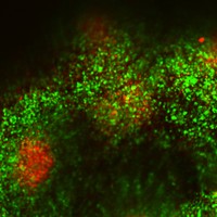
This image shows the interaction between effector T cells (green) and regulatory T cells (red) in a specialized immune structure called Peyer’s patch in the intestine of a mouse. This example of intravital imaging shows how cellular-level events can now be observed inside of animals with microscopes in the UNC-Olympus center
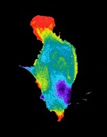
This image shows the activation of a critical signaling molecule, Rac, inside of a living cell from the Hahn lab. This group has pioneered approaches to study protein conformational changes in vivo, including biosensors and image analysis methods for multiplex imaging. Recent work includes genetically encoded approaches to manipulate signaling with light and small molecules.
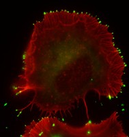
This image shows the localization of the MyosinX protein (green) to the tips of filopodia (finger-like structures shown in red) from Richard Cheney’s lab. This protein helps to transport cargos to the tips of filopodia which are critical for cells to sense their local environment. These structures may also be involved in transmitting viruses from cell-to-cell under certain circumstances.
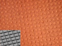
This image illustrates a novel microfabrication technology from the Allbritton lab where cells can be grown on the tops of 3D structures that can later be released using a laser and collected. This allows up to 50,000 cells to screened, while growing in their normal geometry, in a single experiment for properties such as morphology, expression of molecular markers or cell behavior.