Congratulations to Nicholas Pankow, a research specialist in the Pathology Service Core in the Lineberger Comprehensive Cancer Center, for presentation of a poster which was chosen as one of three winners out of 38 posters during the 2023 National Society for Histotechnology conference in Baltimore, Maryland. His poster, titled “P-31: Alternative Strategies For Analyzing Pre-Clinical Mouse Lungs ” was selected “not only for their outstanding content but their ability to effectively communicate using the poster format.” Congratulations to Nicholas and poster co-authors, Gabriela de la Cruz (Director, Pathology Service Core) and Dr. Hannah Atkins, PhD, DVM (Faculty Director, Pathology Service Core)! Read more about the poster competition and other winners on the NSH blog post.
Poster Abstract
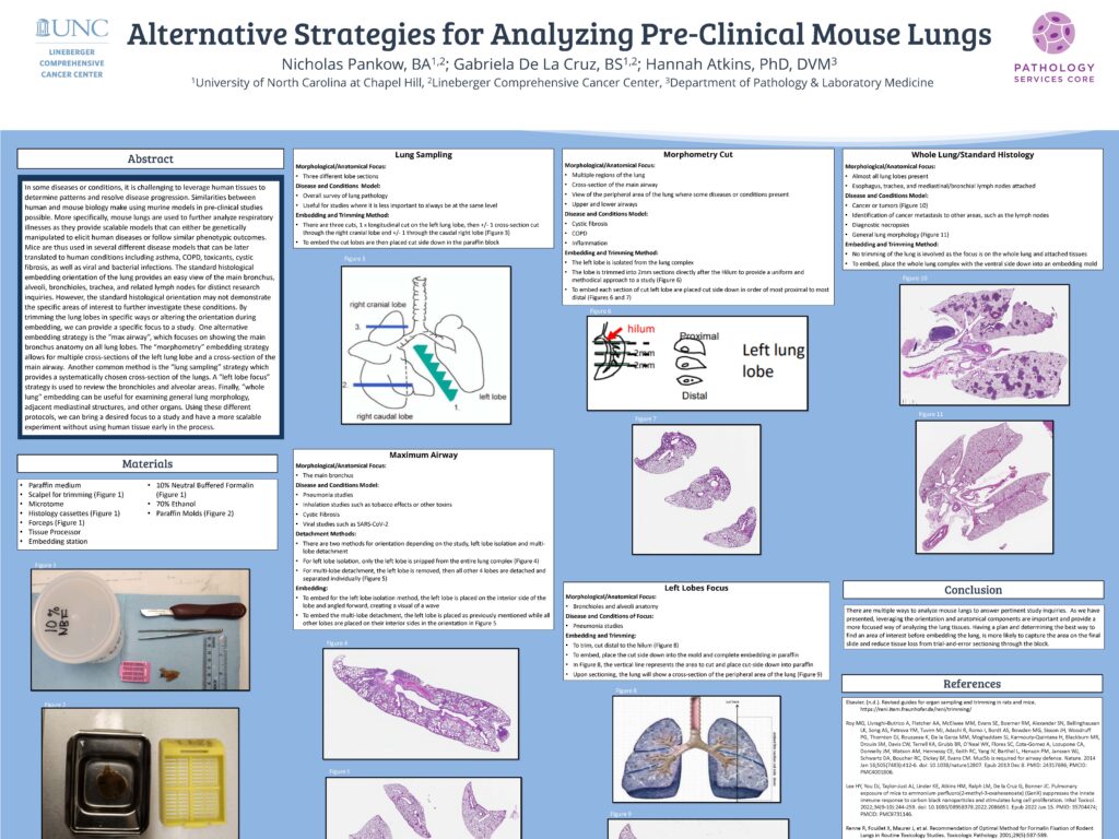
In some diseases or conditions, it is challenging to leverage human tissues to determine patterns and resolve disease progression. Similarities between human and mouse biology make using murine models in pre-clinical studies possible. More specifically, mouse lungs are used to further analyze respiratory illnesses as they provide scalable models that can either be genetically manipulated to elicit human diseases or follow similar phenotypic outcomes. Mice are thus used in several different disease models that can be later translated to human conditions including asthma, COPD, toxicants, cystic fibrosis, as well as viral and bacterial infections. The standard histological embedding orientation of the lung provides an easy view of the main bronchus, alveoli, bronchioles, trachea, and related lymph nodes for distinct research inquiries. However, the standard histological orientation may not demonstrate the specific areas of interest to further investigate these conditions. By trimming the lung lobes in specific ways or altering the orientation during embedding, we can provide a specific focus to a study. One alternative embedding strategy is the “max airway”, which focuses on showing the main bronchus anatomy on all lung lobes. The “morphometry” embedding strategy allows for multiple cross-sections of the left lung lobe and a cross-section of the main airway. Another common method is the “lung sampling” strategy which provides a systematically chosen cross-section of the lungs. A “left lobe focus” strategy is used to review the bronchioles and alveolar areas. Finally, “whole lung” embedding can be useful for examining general lung morphology, adjacent mediastinal structures, and other organs. Using these different protocols, we can bring a desired focus to a study and have a more scalable experiment without using human tissue early in the process.