Gallery
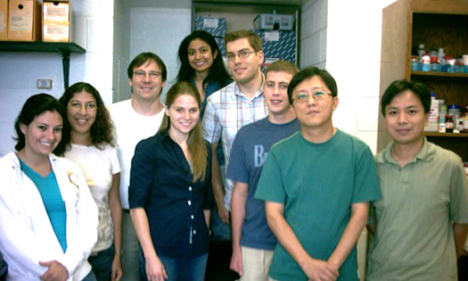
Past and Current Lab Photos
 2007
2007 2008
2008 2010
2010 2011
2011 2013
2013
Kristin's Graduation 2014
2014
Bioinformatics Team 2014
2014 2016
2016 2018
2018
Facilities
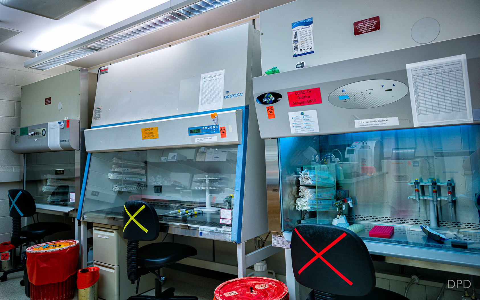
 Setting up 3 COVID research hoods
 Research Technician Brent Eason checks on zika virus-infected mice in a secure ABSL2 isolation area.
Labwork
A time-lapse of KSHV+ HUVEC cells which express GFP, destabilizing after forming tubules on a matrigel plate. Video shows between hours 5-24 after plating cells.
Footage of Dittmer Lab facilities and research. (2020)
Footage of Damania Lab facilities and research. (2020)
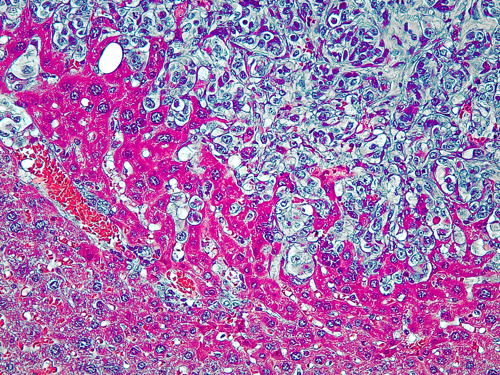
 The image shows a hematoxylin and Eosin (H&E) stain of a liver section belonging to an NSG mouse with metastatic infiltrate. Hepatocytes on lower left of image, with a mass of endothelial cells extravasating into the liver tissue from the upper right. Examining the response of the EC-infiltrated tissue and the overall health of the animal allows us to better understand the clinical implications of KS metastasis and target potential treatments. 200X magnification. This image was chosen as a winner of the 2016 NIH funded research image call. Credit: Imaging/tissue extraction by Anthony B. Eason; lab of Dirk Dittmer, University of North Carolina Lineberger Comprehensive Cancer Center. Mice maintained by UNC Animal Studies Core; staining by UNC Animal Histopathology Core. Research funded by NCI.
 This image depicts HHV-8 Latency-Associated Nuclear Antigen (LANA) expression in an immunohistochemical (IHC) stain of a Kaposi sarcoma (KS) biopsy. The biopsy was obtained from the lymph node of a pediatric patient in Malawi. LANA is identified by dark red staining of the nuclei, and the LANA-negative nuclei are counterstained blue with hematoxylin. The KS cells display a classic spindle cell phenotype, with disruption of the lymph node architecture. Examination of IHC markers from patient samples allows us to correlate findings with clinical data and improve patient outcomes. 200X magnification. This image was chosen as a winner of the 2016 NIH funded research image call. Credit: Imaging and histochemistry by Anthony B. Eason, lab of Dirk Dittmer, University of North Carolina Lineberger Comprehensive Cancer Center. Biopsies provided by Nader Kim El-Mallawany, New York Medical College; and Carrie Kovarik, University of Pennsylvania Research funded by NCI.
 The image depicts a fluorescent multichannel image of the HHV-8 (KHSV)-containing cell line L1T2. Green fluorescein represents HHV-8 LANA, or Latency-Associated Nuclear Antigen, which tethers the KSHV episome to the cellular chromatin (represented by blue DAPI). Red represents actin, stained with rhodamine phalloidin, which highlights the extensive cytoplasm. L1T2 is one of the few cell lines that can retain the KSHV episome in vitro for an extended period of time, while maintaining in vivo tumorigenicity. These cell lines are essential for examining the growth behavior and in vitro drug responses of Kaposi sarcoma. Note the LANA-negative cells, which retain transformation independent of KSHV. 630X magnification. This photo was chosen as a winner of the 2016 NIH funded research image call. Credit: Imaging/staining by Anthony B. Eason; cell line by Sang-Hoon Sin, lab of Dirk Dittmer, University of North Carolina Lineberger Comprehensive Cancer Center. Research funded by NCI.