Hemangiomas
What are these?
 Hemangiomas are non-cancerous (benign) tumors (lmps or masses) made of many thin-walled blood vessels. Hemangiomas may be present at birth (congenital hemangiomas) or may appear days or weeks after birth (infantile hemangiomas). They may occur singly or multiply, may be on the skin (cutaneous) or involve internal organs (such as the liver, lungs, or brain), or both. Although hemangiomas are usually an isolated problem, in some individuals who have large “segmental” (involving a large part or segment of the body, usually both sides of the face or trunk) infantile hemangiomas, they occur as part of a syndrome (a bunch of related problems) called PHACES syndrome. Some individuals may have several different kinds of vascular anomalies (for example, both a vascular malformation and a hemangioma).
How are hemangiomas diagnosed?
Sometimes these are diagnosed in utero at the time of ultrasound when they are usually an incidental finding. Most commonly, the diagnosis of hemangioma can be made just by looking at them. In some cases, special radiologic studies (such as ultrasounds, CAT scans or MRI scans) or even biopsies are needed to be sure of the diagnosis.
What happens to hemangiomas over time?
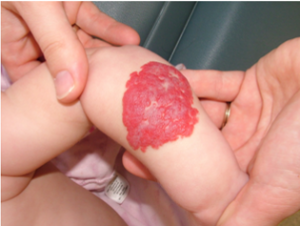
This depends on the type of hemangioma. Infantile hemangiomas are the most common type and are not seen immediately after birth except sometimes as a slight red blush of the involved skin. Within days to weeks, infantile hemangiomas become visible and often grow rapidly. Most infantile hemangiomas stop growing around a baby’s first birthday, and gradually shrink over the next 1-5 years. Often there will be no mark where the hemangioma was. Sometimes, fatty scar tissue will be left where the hemangioma was. Congenital hemangiomas either go away over the first few months of life (RICH, or rapidly involuting congenital hemangiomas) or remain unchanged forever (NICH, or noninvoluting congenital hemangiomas).
What causes hemangiomas?
Although more and more information is being discovered about genes which can be abnormal in some hemangiomas, the cause of most hemangiomas is not known. Infantile hemangiomas occasionally can be seen in more than one family member but usually are not inherited.
How are hemangiomas treated?
Because infantile hemangiomas go away by themselves, they usually do not need to be treated. Reasons for treatment include hemangiomas which are in potentially dangerous places (such as the breathing tube or larynx), which ulcerate and bleed, or which are a cosmetic problem. Treatment, when necessary, should be done by individuals or by multi-disciplinary clinics which are experienced in taking care of children with hemangiomas. Propranolol is a medication which has been used for many years in children for other reasons but which is used more and more for infantile hemangiomas. Laser therapy, sclerotherapy/embolization, or surgical removal sometimes are needed.
What should we look for?
- Where your child’s hemangioma is located will help guide what you should be aware of. For example, in babies with airway hemangiomas, worsening noisy breathing will be important to call your doctor about. A baby with a growing hemangioma of the lip who cannot nurse or suck a bottle should be seen. Your child’s doctors should give you a list of symptoms for which you should call them immediately and others which can wait until normal working hours
- Oozing or bleeding from a hemangioma.
- If a lesion grows unexpectedly, doesn’t go away as predicted, or heals but leaves troubling scar tissue, ask to have your child seen again.
Not all hemangiomas read the text book! Sometimes the original diagnosis needs to be revisited!