The Small Animal Imaging facility will be getting a new PET/CT System in October. The system is funded by a recently awarded NIH high-end instrumentation (HEI) S10 grant (S10-OD023611-01, PI: Zibo Li, PhD). The new system will replace the old GE eXplore Vista PET/CT system, and is expected to have significantly improved performance for PET/CT imaging. (Click Title to read more)
The new system surpasses the old system in all aspects of performance. In particular, several major improvements are described below.
Major enhancements:
– Higher Sensitivity in PET: The SuperArgus PET unit offers doubled sensitivity compared to the old system (8.3 % vs. 4%; central point source sensitivity at 100-700 keV energy window). Higher sensitivity allows researchers to inject less radioactive probes, resulting in lower radiation exposure to animals and researchers, and reduced scan time with similar image quality. For example, acceptable image quality can be achieved by injection of only 30 uCi of Zr-89 witha 5 min acquisition. Dynamic studies can be done with as short as 0.25 s per time frame.
– Large field of view (FOV) in both PET and CT: The new PET unit has an effective axial FOV (length) of 100 mm and transaxial FOV (diameter) of 120 mm, more than double the size of the old system (47 mm in length and 68 mm in diameter). The CT unit also offers a much larger FOV with 80 mm in length and 120 mm in diameter. The system can conduct up to 350 mm long subject by using continuous automated movement of the translation stage. Whole body dynamic scans on mice will be available on the new system too. More attractively, the larger bore size will allow not only small rodents, but also rabbits and guinea pigs for PET and CT imaging. Imaging several mice simultaneously is also possible with the larger bore. These enhanced features will dramatically increase our imaging capacity.
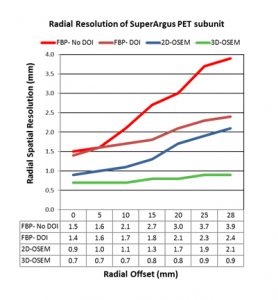
– High Resolution and Fast Reconstruction: The new system can provide sub-millimeter resolution (0.7 mm in center location) when using the 3D iterative OSEM reconstruction algorithm. Better spatial resolution yields less partial volume effects and increases the detection power for small lesions. This will be particularly beneficial for brain imaging on mice, as smaller regions can be differentiated with higher resolution. The PET unit in the new system continues to use the dual-scintillator phoswich detector technology to provide true depth of interaction (DOI) correction. This technology was originally implemented in the eXplore Vista PET/CT system, providing much better resolution at the edges of the FOV. In the new SuperArgus system much improvement has been made in the optical joint between the two crystals, reducing photon loss and enhancing energy resolution. The enhanced DOI technique results in further improvements in radial resolution uniformity, as shown in Figure 2. The implementation of DOI technology not only improves image quality, but also increases throughput by allowing imaging of multiple animals on the same bed, since it is no longer necessary to place each animal in the middle of the bore.
– Real Time PET mode: The new system features a new acquisition mode which uses a time stamp technique to show PET images in real time. Real time PET imaging allows position adjustment during scans, and provides high temporal resolution for monitoring dynamic changes.
– Faster CT and higher CT resolution: The CT unit in the new system offers much faster scans and better image quality. It can be used as a standalone CT system which uses a large flat panel CMOS detector and a micro-focus x-ray source. The highest resolution can be down to 20 micron. Adjustable x-ray energy level ranges from 45 to 110 keV. A standard CT acquisition can be completed within 1 minute. The fast CT scan will dramatically increase imaging throughput, as currently it takes 15 min to conduct a CT scan before PET imaging. Improved CT resolution also enhances image quality, detection power, and diagnosis accuracy in animal studies.
– Enhanced Analysis Tools: The new system comes with the advanced PET image processing software, PMOD, and multimodality fusion software. The PMOD software provides excellent support on dynamic data analysis and parametric mapping. It will provide more standardized and efficient processing of image data analysis, and facilitate many research projects.
Timeline and Planning:
The new PET/CT system is expected to be shipped to the facility in October. About one month will be required for system installation and calibration, followed by acceptance inspection and evaluation. During that time, we will conduct some evaluation studies side-by-side on the new and old systems, the results of which will be reported in our next newsletter. If you are planning some PET studies in October or November, please let us know so we can plan ahead. For new studies, it is recommended to start on the new system. For ongoing studies, we would like to do some comparisons to assess any impact on the study if switching to the new system. Please discuss with us if you have any concerns.