 Joshua Strauss, Ph.D., Director UNC Cryo-EM Core
News contributed by Emily Robinson, Research Technician, UNC Cryo-EM Core
The UNC Chapel Hill School of Medicine’s Cryo Electron Microscopy Core is excited to announce that were awarded North Carolina Biotechnology Center Innovation Impact Grant (GII) for the acquisition of a Thermo-Fischer Scientific (TFS) micro-crystal electron diffraction (microED) package. This package includes an upgraded camera sensor (Ceta D camera, which is a type of CMOS based CCD camera designed specifically for collecting electron diffraction data) and software EPU-D for collecting MicroED data with their existing cryo-transmission electron microscope.
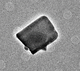
Micro-crystal electron diffraction, or MicroED, is a microscopy method for obtaining atomic resolution structures of proteins and other small molecules. Small, micron sized crystals – for scale, the eye of a needle is about 1,230 microns across – are transferred onto a TEM grid and imaged with the electron microscope under a very weak beam as the sample is continuously rotated. This process takes only a couple of minutes. The images are then analyzed by standard X-ray software programs to generate atomic models. MicroED can be used to determine the 3D structure of different samples, including proteins, drugs, inorganic compounds, zeolites, and polymers that form small micron sized crystals.
This marks an important expansion of UNC’s life science research capabilities. Researchers in academia and industry from a range of disciplines will be able to conduct microED studies on a broad array of specimens. Among other outcomes, this can help researchers better understand and develop new drugs and therapies. With the acquisition of this package, UNC’s Cryo-EM Core will be the first facility in the state to offer MicroED technology to researchers, keeping UNC on the cutting edge of life science technology as well as helping to attract talented students and faculty to the university.
“We are very excited to be able to add microED to the list of imaging method accessible at our core facility and to make this accessible to researchers. This technology is not available in the state of NC and will be a valuable resource for life science researchers in academia and industry, as well as a teaching tool for students.” –Joshua Strauss
The Core expects this package to be a major generator of exciting new work. Researchers can use the microED technique to answer a wide range of research questions, and several research groups have already prepared microcrystals in anticipation of using the Core’s new equipment. Check out their website (med.unc.edu/cryo-em/) to stay up to date with MicroED projects coming out of the core.
Electron micrograph of a micron size biotin crystal and corresponding diffraction image.