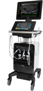
The BRIC Small Animal Imaging (SAI) core facility has recently received a new animal ultrasound imaging system through a NIH S10 shared instrumentation grant (S10-OD034328). The new system, Vevo-F2, will replace the old Vevo-2100 system that has been serving more than 25 research labs in our community. The new system greatly enhances animal ultrasound imaging capability in the SAI facility.
Some new features of the new ultrasound imaging system include:
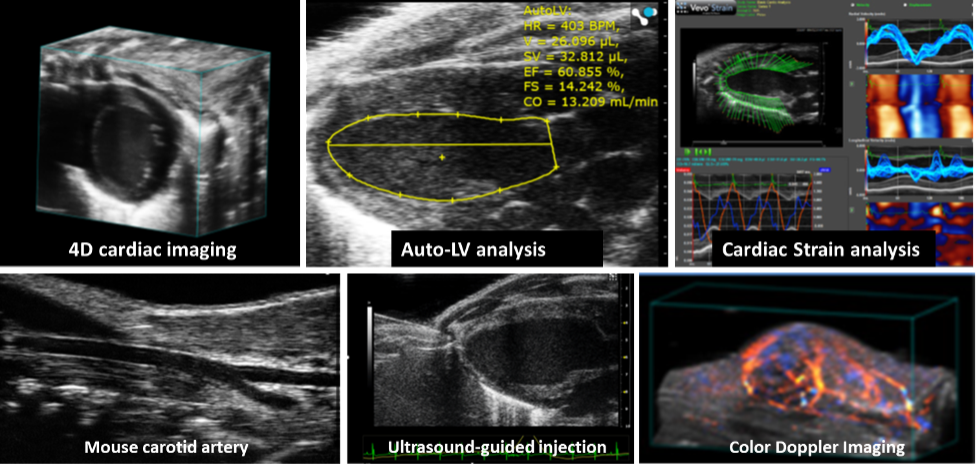
- Six imaging transducers with wide frequency coverage (from 5~ 50 MHz), allowing imaging from mice to small dogs/cats. These will provide either high resolution imaging down to 30 μm, or deep penetration up to 15 mm for larger animals.
- Superior image quality with HD image processing technology, leading to the reduction in speckle noise and artifacts while enhancing tissue contrast
- 4D imaging allowing users to capture dynamic motions in 3D fashion, for example, a complete cardiac cycle in 3D volume.
- Super-fast frame rates (as high as 10,000 frames-per-second) for dynamic acquisition and processing including contrast enhanced imaging;
- Touchpad interface with enhanced workflow
- Three active transfer ports allowing switching between one to another transducer seamlessly
- Additional image analysis packages: Auto-LV (automatic LV analysis in B-mode and Mmode); VevoStrain (cardiac strain analysis); VevoVasc (vascular tissue analysis); VevoCQ (microbubble contrast enhanced image analysis for tissue perfusion).
Some example images of data collected with the Vevo-F2 ultrasound system are shown.
The system has been installed and put in service this November. Please contact the SAI facility at bricasi@med.unc.edu for more information, or visit our website: https://www.med.unc.edu/bric/small-animal-imaging