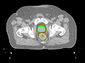
Goal: deform prostate to avoid high dose to the anterior wall of the rectum
Mark Foskey, Bradley C. Davis, Lav Goyal, Sha Chang, Ed Chaney, Nathalie Strehl, Sandrine Tomei, Julian Rosenman, Sarang Joshi
We are studying the use of deformable image registration as a tool in image-guided radiation therapy of the prostate. As modern techniques for conformal therapy attempt to fit the high-dose region as closely as possible to the target volume, it becomes more essential to ensure that the tumor is in the expected location, and to assess the dose actually delivered to each organ.
We use deformable image registration in two ways. First, we use it for automatic segmentation of images acquired at treatment time, by deforming manual segmentations of the planning day image. Second, we use it to estimate the dose actually delivered, by deforming calculated dose distributions from each treatment day. In each case, the deformations we apply are computed by registering a treatment image to a planning image.
We perform our registration in two stages. We first perform a translation based on bony landmarks, and then a deformable registration in a region of interest. The images to the right illustrate the effects of the stages. Only the conturs differ; the images are all the same slice from the planning CT of a patient. The red contour is the location of the prostate in the planning CT, and the yellow contour is the rectum. The green contours indicate the locations of the prostate in the daily images. The top image shows the prostate locations as treated. The middle images shows the prostate locations after translation to align bony landmarks. And the bottom image shows the locations of the prostate after deformable registration is performed. In the latter case, the image deformation was applied to 3D prostate models derived from the manual segmentations, which were then resliced to get the deformed contours.

Average radial distance between
deformed planning segmentation
and 2 rators.
Selected Publications
- Foskey M, Davis B, Goyal L, Chang S, Chaney E, Strehl N, Tomei S, Rosenman J, and Joshi S. 2005. Large deformation 3D image registration in image-guided radiation therapy. Under Review.
- Davis B, Foskey M, Rosenman J, Goyal L, Chang S, and Joshi S. 2005. Automatic segmentation of intra-treatment CT images for adaptive radiation therapy of the prostate. Proc. Medical Image Computing and Computer-Assisted Intervention. To appear.
- Foskey M, Rosenman J, Goyal L, Chang S, and Joshi S. Calculating biological effective dose in the presence of organ deformation. AAPM 2005.