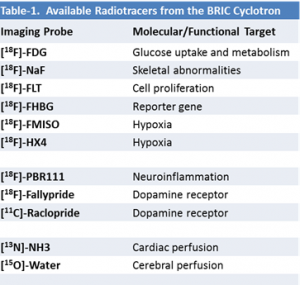
The BRIC Cyclotron Facility has expanded its menu of radiolabeled tracers available for PET imaging. More than ten PET probes labeled with F-18, C-11, N-13, and O-15 radioisotopes can now be requested from the facility for neurological, cardiovascular, or cancer research studies.
We are proud to announce that the BRIC Cyclotron and radiochemistry facility is now capable to provide more PET imaging probes beyond the commonly available 18F-FDG agent. More than ten PET tracers have been added to the radiotracer menu that are now available to all investigators (Table-1). The expanded menu will greatly enhance our molecular imaging capability and facilitate many cancer and neuroscience research.
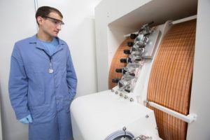
The BRIC cyclotron facility is equipped with a GE 16.5MeV PETtrace cyclotron, hot cells and minicells for radiotracer synthesis. In addition, the core also has a GMP level clean room, two dispensing cells, and QC setup to provide probes for human imaging research and clinical trials. The newly installed cyclotron has 6 targets which can be used to produce various radioisotopes including F-18 (two targets), C-11(one target), O-15 (one target), N-13 (one target), and one target reserved for future radio-metal production. Under the core Director, Dr. Zibo Li, the facility has made tremendous progress since the installation of the cyclotron facility. Many new imaging probes are now available for preclinical imaging research, including F-18 labeled FLT (tumor proliferation), FMISO (tissue hypoxia), HX4 (tissue hypoxia), fallypride (dopamine receptor imaging), and PBR111 (neuroinflammation), C-11 labeled raclopride (dopamine receptor imaging) N-13 labeled ammonium (cardiac perfusion), and O-15 labeled water (cerebral blood flow).
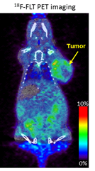
18F-FLT has been used widely in oncology for tumor proliferation [1]. It has been reported that it is superior to FDG for detecting recurrent high grade brain tumors [2], and have been used to predict treatment outcome. An example of PET imaging with 18F-FLT is shown in Figure-1. 18F-FMISO and 18F-HX4 are both targeting tumor hypoxia [3, 4]. 18F-FMISO is the mostly used hypoxia imaging marker with several clinical trials for head and neck cancer, lung cancer, cervical cancer, and brain tumors. An image example is shown in Figure 2. 18F-HX4 is more hydrophilic compared to FMISO, resulting faster blood clearance and less waiting time after injection. Along with 18F-FDG, the core can provide comprehensive molecular imaging probes for cancer research.

Fig. 2. Tumor hypoxia study using 18F-FMISO PET imaging and autoradiography imaging compared to hypoxia immunostaining image.
18F-PBR111 is a relatively new PET probes with high affinity and selectivity for the translocator protein (TSPO) expressed on activated glia cells. It has been used in several neuroinflammation animal models [5]. Figure 3 shows an example of using PBR111 to image neuroinflammatory mouse model. Fallypride and raclopride both are widely used PET tracers for imaging dopamine D2 receptors with F-18 or C-11 radiolabeling [6]. They can be used to monitor changes on Dopamine D2 receptor related diseases such as Parkinson, drug addiction, and epilepsy study [7].


Fig. 4. Serial axial frames from PET imaging with 13N-ammonia to study cardiac perfusion on a rat model.
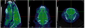
Fig. 5. PET imaging with 15O-water on a rat model for cerebral blood perfusion application.
Dr. Zibo Li’s group also has the capability to design and manufacture custom tracers for your study. The probe development team will work together with investigators to customize imaging probes targeting your molecular event. Please contact Dr. Zibo Li for more information. Infrastructure to provide these probes for human imaging study is undertaking. Please contact Dr. Eric Smith for any regulations on human imaging study with these probes.
References:
1. Peck M, Pollack HA, Friesen A, Muzi M, Shoner SC, Shankland EG, et al. Applications of PET imaging with the proliferation marker [18F]-FLT. Q J Nucl Med Mol Imaging. 2015;59(1):95-104. Epub 2015/03/05.
2. Chen W, Cloughesy T, Kamdar N, Satyamurthy N, Bergsneider M, Liau L, et al. Imaging proliferation in brain tumors with 18F-FLT PET: comparison with 18F-FDG. J Nucl Med. 2005;46(6):945-52. Epub 2005/06/07.
3. Lee ST, Scott AM. Hypoxia positron emission tomography imaging with 18f-fluoromisonidazole. SeminNuclMed. 2007;37(6):451-61.
4. Dubois LJ, Lieuwes NG, Janssen MH, Peeters WJ, Windhorst AD, Walsh JC, et al. Preclinical evaluation and validation of [18F]HX4, a promising hypoxia marker for PET imaging. Proc Natl Acad Sci U S A. 2011;108(35):14620-5. Epub 2011/08/30.
5. Colasanti A, Guo Q, Muhlert N, Giannetti P, Onega M, Newbould RD, et al. In Vivo Assessment of Brain White Matter Inflammation in Multiple Sclerosis with (18)F-PBR111 PET. J Nucl Med. 2014;55(7):1112-8. Epub 2014/06/07.
6. Mukherjee J, Yang ZY, Brown T, Roemer J, Cooper M. 18F-desmethoxyfallypride: a fluorine-18 labeled radiotracer with properties similar to carbon-11 raclopride for PET imaging studies of dopamine D2 receptors. Life Sci. 1996;59(8):669-78. Epub 1996/01/01.
7. Virdee K, Cumming P, Caprioli D, Jupp B, Rominger A, Aigbirhio FI, et al. Applications of positron emission tomography in animal models of neurological and neuropsychiatric disorders. Neuroscience and biobehavioral reviews. 2012;36(4):1188-216. Epub 2012/02/22.
(All the images in the figures were taken on the GE eXplore Vista PET/CT system in the SAI facility)