High resolution CT imaging is used to delineate cardiac structure of Xenopus froglets in Frank Conlon’s group.
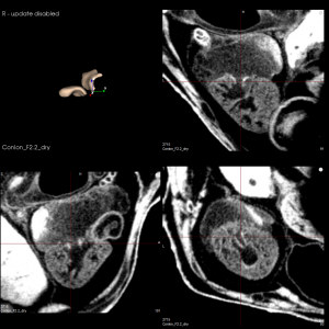
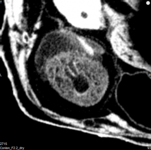
We have recently developed a high resolution contrast enhanced CT imaging protocol for a study on Xenopus froglets. Dr. Frank Conlon’s group has been using Xenopus froglets as an animal model to study the genetic causes of congenital heart disease. The focus of the study is to ascertain how the Xenopus heart develops and grows and to help determine its structural similarity to the mammalian heart. Using high resolution microCT, detailed structures and defects in froglet heart can be delineated without standard histological methods on the heart tissue. The froglet’s were fixed at NF stage 66 and had a torso length of approximately 1cm, which is a newly metamorphosed froglet. The images below were obtained with the SCANCO uCT40 with a voxel size of 10um following PTA contrast staining. Post-processing was completed with VivoQuant software. The image below shows a 3D rendering of the froglet heart with the segmented valves shown in light pink.
