Histology Core
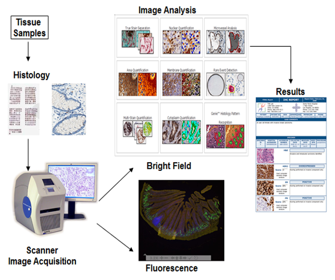
The Cell Services & Histology Core provides a full range of histology services, encompassing routine and specialized grossing, tissue processing, paraffin embedding, microtomy, H&E and special staining, and immunohistochemistry. Consultations are also available. The core also offers image analysis through the Translational Pathology Laboratory (see figure). These services are available to CGIBD members and non-members.
Contact:
Tope Keku, PhD (Director)
tokeku@med.unc.edu
Nikki McCoy
amber_mccoy@med.unc.edu
SERVICES AND FEES:
| Services | CGIBD Member | Internal Non-Member | External (Academic) | External (Corporate) |
|---|---|---|---|---|
| Standard Package (P&E, 1 Section, 1 H&E)/cassette | $15.81 | $22.63 | $29.44 | $52.44 |
| Paraffin Processing | $4.00 | $5.11 | $6.22 | $10.22 |
| Grossing | $8.00 | $10.00 | $12.00 | $20.00 |
| Paraffin Embedding | $4.00 | $5.11 | $6.22 | $10.22 |
| Re-embedding | $3.00 | $3.84 | $4.67 | $7.67 |
| Additional Pieces per Block | $4.00 | $5.11 | $6.22 | $10.22 |
| Section (serial or skips) | $3.50 | $5.25 | $7.00 | $12.00 |
| H&E Stain | $4.50 | $7.25 | $10.00 | $20.00 |
| Trichrome Stain | $12.21 | $21.11 | $30.00 | $55.00 |
| PAS-H Stain | $8.00 | $14.00 | $20.00 | $40.00 |
| Von Kossa Stain | $8.00 | $14.00 | $20.00 | $40.00 |
| Other Stains* | $8.00 | $14.00 | $20.00 | $40.00 |
| Digital Slide Scanning | $6.00 | $10.00 | $14.00 | $23.00 |
| Slide Box - 25 place | $9.00 | $11.50 | $14.00 | $20.00 |
| Slide Box - 100 place | $13.00 | $16.61 | $20.22 | $30.00 |
| Rush Order* | $35.00 | $45.00 | $55.00 | $90.00 |
| Staff hourly rate: Experimental Design | $75.00 | $97.50 | $120.00 | $160.00 |
| Additional Services | TBD | TBD | TBD | TBD |
LARGE FORMAT/cassette sample
SUPER MEGA BLOCK SECTIONING
| Services | CGIBD Member | Internal Non-Member | External (Academic) | External (Corporate) |
|---|---|---|---|---|
| Paraffin Processing | $16.00 | $20.50 | $25.00 | $38.00 |
| Paraffin Embedding | $16.00 | $20.50 | $25.00 | $38.00 |
| Sections (serial or skips) | $9.00 | $12.00 | $15.00 | $20.0 |
| H&E Stain | $14.00 | $19.50 | $25.00 | $29.00 |
| Trichrome Stain | $22.00 | $33.50 | $45.00 | $65.00 |
| Large format Package (P&E, 1 Section, 1 H&E) | $55.00 | $70.00 | $85.00 | $125.00 |
Depends on the costs of reagents.
Rush orders must be discussed first before request is made.
Image analysis
Digitized images can be accessed through a secure website. The investigators will have access to Aperio Imagescope software for annotation and scoring of images. Scanned images in the Spectrum Plus database can be shared with colleagues and viewed simultaneously. Investigators can request full (edit and annotate) or read only (read only) access for collaborators and others.
Microscopes
The histology core has a Zeiss AX10 Imager and Zeiss AX10 Primo Star Microscope and an Olympus 1X81 inverted fluorescence microscope.
