
Dr. Jonathan Fay, in collaboration with Jared Peck and Dr. Joshua Strauss of the UNC CH CryoEM Core, published a method paper in ICUrJ (International Union of Crystallography Journal) outlining their success in optimizing the function of a microscope for cryogenic electron microscopy (CryoEM). CryoEM is an imaging technique that yields 3D structures of large macromolecular complexes, viruses, and cells in order to provide atomic insight into the biological processes underlying life itself.
The goal of the project was to develop procedures to maximize the throughput and performance of UNC-CH’s cryogenic transmission electron microscope (cryoTEM), called a Talos Arctica. This model of electron microscope is still considered by some to be a “screening microscope” and therefore less capable of high-speed, high-resolution data collection. However, the team saw that it had potential to be used to quickly and efficiently collect quality images.
In the optimization process, the team evaluated the effects of different camera and microscope parameters on the imaging of a protein called apoferritin (prepared by Dr. Nathan Nicely of the UNC-CH Protein Expression Core). They found that
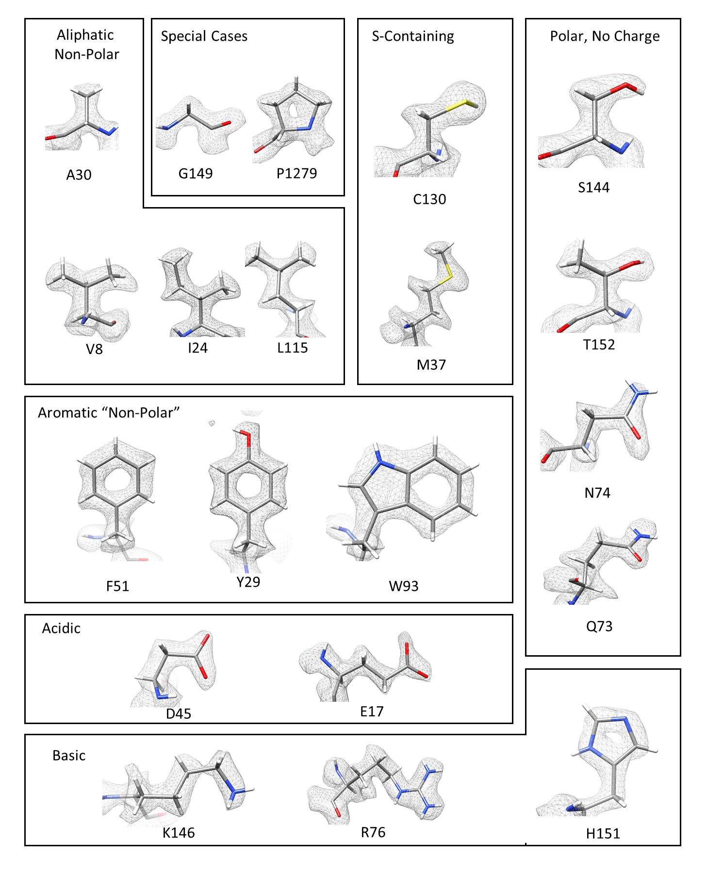
with software adjustments, it is possible to use the Talos Arctica to collect data at a rate of 720 “movies” per hour (roughly 17,000 per day). The movies were subsequently used to generate high-resolution (1.8-2.2 Å) maps of apoferritin’s structure. The broad impact of this work is that the adjustments made on the Talos Arctica can also be applied to other models of electron microscopes, and thus can expedite data collection for many in the field of cryoEM. The improvements will also directly benefit those using UNC-CH’s CryoEM Core facility.
Core Director and author Dr. Joshua Strauss says about the work that “[the team] believes improving data collection speed while maintaining data integrity is an emerging trend in the field of cryo-EM. Procedures for high-speed data collection and benchmarks for data quality validation that are microscope and camera specific should be present in the literature, as they are critical in improving technical understanding of cryo-EM.”
This project was funded by the UNC Core Facilities Advisory Committee’s voucher program with the specific goal to expedite sample quality assessments for structural studies of GPCRs, a large group of signaling proteins that includes many therapeutic targets with unknown structures. The team plans to implement their new tools to make a dent in this pool of unknowns.
Author Dr. Jonathan Fay said, “With the vetted methodological improvements we have put into practice at the UNC CryoEM Core, it is now routine to collect data for around three protein samples in a 16-hour time period, with each data set typically resulting in nominal resolutions of around 2.5-3 Ångstroms.” He noted that this resolution was achieved for a non-symmetrical membrane protein complex. Having detailed structural data on GPCRs will allow researchers to better understand and control signaling pathways that lead to disease.
The Cryo-EM core thanks Anna Wheless from Dr. Saskia Neher’s lab for taking the lab photos of the investigators and writing up this news story.