Image Analysis
How we will help you
At MSL we will maximize your chances of success by:
- Consulting with you about your image analysis options.
- Developing optimal image analysis workflows in FIJI, Imaris, SyGlass and Autoquant. This includes writing macros to automate analysis steps.
- Training you to execute those workflows. We can record training sessions so you can review them afterwards.
Please contact us as early as possible in your project.
Analysis workstation – “Minerva”
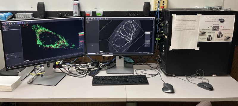
A powerful computer on which the software described below is installed, which can be used in person or remotely. We provide temporary storage for data being analyzed on this workstation and on our dedicated server. Note that VR software (Syglass) and hardware can only be used in person.
Calendar Usage instructions Remote access instructions MSL server instructions
CPU: 2 x Intel Xeon E5-2667 V4 3.2 GHz Eight Core
RAM: 512 GB
GPU: NIVIDIA PNY Quadro M6000 PCI-E 24 GB
Storage:
- 2 x Intel 750 1.2 TB PCI-E SSD
- 8 x Sabrent Rocket Q 8TB NVMe PCIe M.2 in RAID 10 (around 30TB of available storage)
- HighPoint SSD7140 PCIe 3.0 x16 8-Channel M.2 NVMe RAID Controller
- IcyDock Tough Armor 4 Bay 2.5″ SATA HDD & SSD for hot swap drives. More on this option here.
Network: Intel Converged 10 Gb Network Adapter X540-T1
Virtual reality: HP Reverb G2 Headset
Mouse: Logitech MX Master with 2 scroll wheels (used for indpendent zooming and slicing functions in Imaris)
Software
We provide support in the following four softwares packages and can install other software on our analysis workstation if needed (MATLAB, Python, cellpose, CellProfiler, QuPath, etc):
Multidimensional visualization and analysis software
Free Imaris Viewer can be used for visualization and snapshots.
Full version available at MSL can be used for visualization, snapshots, movies and analysis.
Deconvolution of 3D datasets
MSL's Autoquant tutorialVirtual reality and analysis software
Free SyGlass View allows visualization (VR headset required).
Full version available at MSL can be used for visualization, snapshots, movies and analysis.
Free, open-source software for image analysis
Download FIJI MSL's Basic FIJI tutorial Image.sc community forumCollaborations with our users
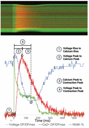
Voltage, calcium ions and contraction kinetics measured simultaneously, at 0.8kHz, in beating cardiomyocytes
- Data acquired on our Zeiss LSM900 laser scanning confocal.
- Collaboration with Ike Emerson, in Frank Conlon’s lab.
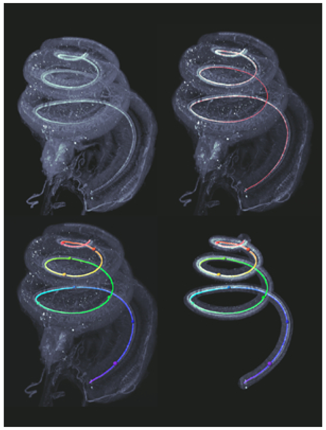
Three-dimensional frequency maps and cell counts in the cochlea, using Imaris.
- Data acquired on our Lavision UltraMicroscope II light-sheet system.
- Collaboration with Ken Hutson, in Doug Fitzpatrick’s lab.

Quantification of arterial injury in the rat carotid artery, using Imaris.
- Data acquired on our Lavision UltraMicroscope II light-sheet system.
- Collaboration with Nick Buglak, in Ed Bahnson’s lab.
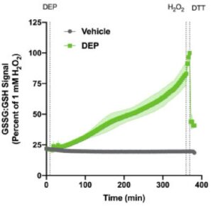
Measurement of hydrogen peroxide and glutathione oxidation in live cells, using a fluorescent reporter, analyzed with FIJI macro.
- Data acquired on our IX81 microscope.
- Collaboration with Sam Faber, in Shan McCullough’s lab.

Measurement of precise 3D distances between DNA loci, using Imaris.
- Data acquired on our BX61 microscope.
- Collaboration with Meghan Schertzer, in Mauro Calabrese’s lab.
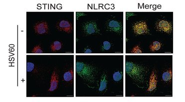
Quantification of subcellular colocalization in cell culture, using FIJI and Imaris.
- Data acquired on our Zeiss LSM700
- Collaboration with Alex Petrucelli, in Jenny Ting’s lab.