Cataract & Other Surgical Curricula

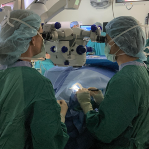
Residents perform their first 50 cataract surgeries during this rotation. UNC Ophthalmology’s unique Cataract Clinic UNC Hospitals Hillsborough campus allows residents to take excellent of surgical patients from the first surgical evaluation through the postoperative period. By the end of this course, residents are able to perform entire surgeries independently, and learn to manage complex cases and surgical complications. After this rotation, residents rotate immediately at the Fayetteville VA, allowing them to build on this initial surgical experience.
Cataract Surgical Curriculum Pterygium Surgical Curriculum Corneal Topography