High resolution and high tissue contrast CT imaging on tissue specimen is being performed at the SAI facility using new tissue preparation protocols.
Traditional study of cardiovascular defects in mouse embryos relies on histological analysis on tissue sections, which is limited to selected sections with possibility of missing critical regions of interest, and is prone to cutting artifacts. Although 3D reconstruction on serial sections has been developed, it is highly labor-intensive and has the same limitation of cutting artifacts. Computer tomography (CT) imaging provides high resolution 3D volumetric information without damaging organs or tissues of interest, and can delineate internal structures as small as 2 um using a preclinical high resolution CT scanner. However, one major limitation with CT imaging is its low tissue contrast due to the similar x-ray attenuation properties from different soft tissue types. To improve soft tissue contrast, CT contrast agents have been introduced to provide better imaging capability. Specifically, preserved specimens, such as fetus or other soft tissue specimens, will be stained with certain CT contrast agents before imaging, and high resolution CT imaging will be performed on the processed tissue in high tissue contrast fashion. Here we would like to introduce the utility of using contrast enhanced CT to obtain high resolution and high contrast 3D images of tissue specimens.
Several contrast agents used to prepare tissue specimens for CT imaging include osmium tetroxide [1], inorganic and organic iodine [2], and phosphotungstic acid (PTA) [3]. The common requirements for these contrast agents are: 1) Their capability of high x-ray attenuation property; 2) Their ability to penetrate into fixed tissue specimens. Inorganic iodine agents such as Lugol solution can rapidly diffuse into fixed tissue and can complete staining on most mouse specimens after soaking overnight. It works well even for tissue with prolonged fixation time in formalin [4]. PTA is a much larger molecule (2.8 KD) with slower penetration. Whole tissue penetration takes a little longer time. One of the advantages for PTA staining is its ability to bind to proteins including collagen, and can offer good tissue contrast based on different binding levels in different tissue types. Osmium tetroxide, although used widely in electronic scanning and was introduced first for ex-vivo CT imaging compared to other agents , has been used much less now largely due to its acute toxicity. Table-1 below lists the common CT contrast agents for ex-vivo CT imaging.
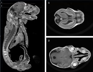
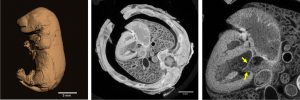
The SAI facility has developed several contrast preparation protocols for CT imaging on various specimens, including mouse and rat fetus, froglets, and partial body specimens. We have demonstrated the broad applicability of contrast enhanced CT on animal specimens with high resolution and high tissue contrast. Figure 1 and 2 are PTA stained rat fetus with high resolution CT which are used to study the cardiac and brain morphology in a toxicity study. Figure 3 shows the Lugol stained floglet in a study to depict the size of heart valves in different disease models. Such imaging method offer great utility for the nondestructive visualization and characterization of small animal specimens, and opens a wide application in comparative, developmental and morphological research on animal models.
Table -1: Common contrast agents for postmortem CT imaging
|
Name |
Main ingredients |
Characteristics |
|
Lugol |
Aqueous potassium triiodide |
Easy penetration and preparation, good contrast |
|
I2E, I2M |
1% Iodine in 100% ethanol (I2E) or methanol (I2M) |
Easy penetration, good contrast |
|
PTA |
1% (w/v) phosphotungstic acid |
Large molecular with slowed penetration, high contrast |
|
Osmium |
Osmium tetroxide |
Good contrast, high toxicity |

Figure 3. Images of Xenopus froglet heart after treatment with Lugol staining methods. Top left: heart valve shown in volume rendered image; Other panels: different views of froglet heart.
For more information on the tissue preparation and imaging protocol details, please contact the facility, bricsai@med.unc.edu, or call 919-966-2855.
References:
[1] Johnson JT, Hansen MS, Wu I, Healy LJ, Johnson CR, Jones GM, et al. Virtual histology of transgenic mouse embryos for high-throughput phenotyping. PLoS Genet 2006;2:e61.
[3] Dunmore-Buyze PJ, Tate E, Xiang FL, Detombe SA, Nong Z, Pickering JG, et al. Three-dimensional imaging of the mouse heart and vasculature using micro-CT and whole-body perfusion of iodine or phosphotungstic acid. Contrast media & molecular imaging 2014;9:383-90.
[4] Metscher BD. MicroCT for comparative morphology: simple staining methods allow high-contrast 3D imaging of diverse non-mineralized animal tissues. BMC physiology 2009;9:11.
|
|