Pharmacology’s Wesley Legant, PhD, and Department of Biomedical Engineering’s, Yu Shi, PhD, latest paper, “Smart Lattice Light-Sheet Microscopy for Imaging Rare and Complete Cellular Events” was featured on the cover of Nature Methods!
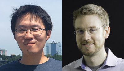
(Above L): First author, Yu Shi, PhD, Postdoctoral Researcher, Legant Lab; (Above R): Senior author, Wesley Legant, PhD, Assistant Professor.
Abstract
“Light-sheet microscopes enable rapid high-resolution imaging of biological specimens; however, biological processes span spatiotemporal scales. Moreover, long-term phenotypes are often instigated by rare or fleeting biological events that are difficult to capture with a single imaging modality. Here, to overcome this limitation, we present smartLLSM, a microscope that incorporates artificial intelligence-based instrument control to autonomously switch between epifluorescent inverted imaging and lattice light-sheet microscopy (LLSM). We apply this approach to two unique processes: cell division and immune synapse formation. In each context, smartLLSM provides population-level statistics across thousands of cells and autonomously captures multicolor three-dimensional datasets or four-dimensional time-lapse movies of rare events at rates that dramatically exceed human capabilities. From this, we quantify the effects of Taxol dose on spindle structure and kinetochore dynamics in dividing cells and of antigen strength on cytotoxic T lymphocyte engagement and lytic granule polarization at the immune synapse. Overall, smartLLSM efficiently detects rare events within heterogeneous cell populations and records these processes with high spatiotemporal four-dimensional imaging over statistically significant replicates.”
~The above abstract is an excerpt from the Nature Methods, 21, pages 301–310 (2024) article published by the Legant Jan. 2, 2024.
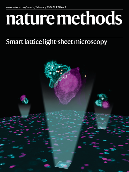
The image to the left shows immune synapses formed between cytotoxic T lymphocytes (cyan) and tumor cells (magenta) within a population of cultured cells. Cytotoxic granules are shown in yellow.
~Cover for the Feb 20, 2024 issue of Nature Methods. Image by Yu Shi and Wesley Legant; Cover design by Thomas Phillips.
Other co-authors on the paper are: Jimmy S. Talbot, Daniel E. Milkie, Timothy A. Daugird, Chelsea Q. Yang, Alex T. Ritter, and Andrea Giovannucci.
Congrats to all!