Urinary Stones
Page Contents
What is Stone Disease?
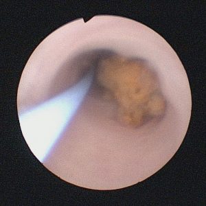
Urinary calculi, solid particles in the kidneys, bladder, or ureter, are of various chemical compositions — calcium oxalate, calcium phosphate, uric acid, cystine, and struvite.
Renal calculus disease is one of the most ancient afflictions of mankind and remains a common cause for both office and emergency room urologist’s visits. In the United States, approximately one in eight males and one in 15 females will form a urinary tract stone within their lifetime.
After an initial episode of renal calculus, the recurrence rate of stones within these patients is as high as 50% within the next five years. Men and patients with a family history of stone disease are three-fold more likely to have a stone than the general population. However, the gender distribution in stone disease is shifting with newer studies showing an increased stone prevalence for females almost equal to males. The Southeast portion of the country, including North Carolina, is considered the Stone Belt because of its highest prominence in stone disease. Researchers have questioned if this increased occurrence may be due to differences in temperature, sunlight, and fluid consumption.
Symptoms
(Read More)Treatment
The majority of stones in the kidney or in the ureter may be observed conservatively for a period of time if they are not causing pain, blood in the urine, infection or blockage of the kidney. Many stones in the ureter, particularly those 4 to 5 mm or less are likely to pass spontaneously through the urine tract though it may take several weeks for this to occur. Some medications such as an alpha-blocker may be given to aid in the passing of a stone.
For patients with larger stones or stones causing symptoms such as pain, blood in urines/infection or blockage of the kidney, surgical treatment may be necessary.
With the development of shock wave lithotripsy (SWL), small caliber endoscopes and safe percutaneous access to the upper urinary tract, the entire urinary tract can be assessed, visualized and treated with minimally invasive techniques. While treatment options continue to improve, many patients require advanced surgical techniques for difficult stone management issues caused by stone type, location, size, and patient anatomical differences.
Shock Wave Lithotripsy (SWL)
(Read More)Shock Wave Lithotripsy (SWL) is a minimally invasive treatment that was introduced in 1980 after years of research between Dornier, Inc. and the University of Munich. This technology uses shock waves generated outside the body by a lithotripter and are then targeted by fluoroscopy or ultrasound to fragment stones within the urinary tract. Fragmentation occurs through tensile stress that removes surface material and pulverization of the stone through the application of multiple shock waves. The number of shock waves required for adequate stone fragmentation depends on the composition and size of the stone, the focal pressure, energy density, and fluid interface. Stones that fragment easily include calcium oxalate dihydrate, uric acid, and struvite. Stones that are difficult to fragment include calcium oxalate monohydrate, cystine, and calcium phosphate. The use of shock wave lithotripsy is dependent on the size, position, and anatomic features of the stone and is less effective with large stones and in obese patients due to difficulty in getting the stone into the focal point. Once a stone is adequately treated, the fragments can be passed spontaneously from the urinary tract.
Ureteroscopy
(Read More)
Rigid ureteroscopy has been used since the 1980s and was initially indicated for the management of distal ureteral stones. The development of smaller, semi-rigid ureteroscopes and more recently, flexible deflectable ureterorenoscopes, allows routine endoscopic evaluation of the entire urinary collecting system. Both rigid and flexible ureteroscopy are used for stone diagnosis and treatment and allows for complete stone removal at the time of surgery. Ureteroscopy can be indicated for any stone located throughout the urinary system. Small stones in the lower ureter, can be extracted by a basket or forceps passed through a rigid scope that has been passed over a working guidewire or alongside a safety guidewire. Larger ureteral calculi and stones within the kidney can be treated with a flexible scope electrohydraulic or Holmium laster intracorporeal lithotripsy to fragment the stone(s) prior to passage or removal. A temporary ureteral stent is often placed at the end of the surgery to facilitate drainage for a few days after surgery. Ureteroscopy can be used for other medical conditions such as to investigate gross hematuria and positive urine cytology, fulguration of epithelial tumors and management of ureteral strictures, obstructed calyces, and ureteropelvic junction(UPJ) obstruction.
Percutaneous Nephrolithotomy
(Read More)Endoscopic or intracorporeal management of stones through a percutaneous tract into the renal collecting system is called percutaneous nephrolithotomy (PNL). This technique was developed in 1975 by Fernstrom and Johanson. It can be used for most renal and upper ureteral stones (such as stones within the lower pole calyx, with a calyceal diverticulum or a staghorn calculi) but is used mostly for large stones, greater than 2 cm or 1.5 cm in the lower pole that are not easily managed by ESWL or ureteroscopy, and as a salvage procedure for failed ESWL. PNL is performed under general anesthesia. The patient is placed in the prone position. Needle access to the kidney is obtained through the back and then a guidewire is then passed down the ureter and the tract is dilated with a balloon dilator. A hollow plastic sheath is placed through the tract and then a rigid or flexible nephroscope is passed. Stones less than 1 cm in size can be manually extracted through the plastic sheath using grasping forceps. If the stones are larger than 1 cm, intracorporeal lithotripsy using ultrasonic, electrohydraulic or laser lithotripsy is performed. (PNL)
Open, Robotic, and Laparoscopic Lithotomy
(Read More)With our current endoscopic treatments, only 1% to 5% of stones require a more invasive procedure for removal. Very select patients with abnormal kidney anatomy such as a horseshoe kidney, pelvic stenosis, a nonfunctional kidney, or additional kidney abnormalities such as a concurrent ureteropelvic junction obstruction may be candidates for more invasive treatment including open, robotic or laparoscopic lithotomy procedures.
Prevention
Given the high recurrence rate in stone disease, development of preventive strategies to minimize recurrence is almost as important as the treatment of an individual stone. Patient metabolic abnormalities for stone risk can be found in up to 90% of stone formers.
(Read More)Any patient who has had at least one stone should have a few basic tests including serum labs to check for systemic causes of stones and a stone analysis to determine stone composition. Patients with recurrent stones or those at high risk for recurrence should perform a 24-hour urine test (Litholink) to tailor preventive strategies to their individual risks. Patients at high risk include any child with stones (<18 years old), patients with only one (functional) kidney, patients with abnormal kidney anatomy, and patients with a history of gastrointestinal diseases including Crohn’s Disease, ulcerative colitis, bowel resection, gastric bypass and other bariatric surgeries. These patients may also benefit from an additional evaluation in the Multidisciplinary Stone Clinic with our nephrologist (medical kidney disease) and nutritionist to maximize stone prevention.
Because many risk factors for kidney stones are diet related, nutritional changes are the cornerstone of any stone prevention strategy. Stone patients should increase fluid intake to 2 to 2.5 liters per day. Additionally, other common recommendations include a low salt diet, moderate calcium intake, low animal protein diet, low oxalate diet, and increased dietary citrate.
Medication therapy such as thiazide diuretics and alkalinization medications may also be used for stone prevention. Generally, these are started following a trial of dietary changes in high-risk patients or those with stone recurrence based on persistent risk factors noted on 24-hour urine testing.
Conclusion
First line therapy for urinary stones typically involves minimally invasive surgical procedures for obstructing stones that cause symptoms and do not pass spontaneously in a reasonable time. Treatment decisions are based on the suspected stone type, size, location, renal anatomy, and renal function. Morbidity, hospitalization, and cost are often reduced significantly with minimally invasive treatments such as extracorporeal shock wave lithotripsy, ureteroscopy, and percutaneous nephrolithotripsy. More invasive surgical treatments are rare but indicated in select cases. Patients recover more quickly and have a quicker return to normal activity with the less-invasive surgical options that are available. Because of the high rate of recurrence in stone patients, optimizing stone prevention strategies is as important as treatment of individual stones.
