Understanding how proteins bend, twist, and shape-shift as they go about their work in cells is enormously important for understanding normal biology and diseases. But a deep understanding of protein dynamics has generally been elusive due to the lack of good imaging methods of proteins at work. Now, for the first time, scientists at the UNC School of Medicine have invented a method that could enable this field to take a great leap forward.
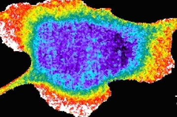
The scientists’ new “binder-tag” technique, described in a paper in Cell, allows researchers to pinpoint and track proteins that are in a desired shape or “conformation,” and to do so in real time inside living cells. The scientists demonstrated the technique in, essentially, movies that track the active version of an important signaling protein – a molecule, in this case, important for cell growth.
“No one has been able to develop a method that can do, in such a generalizable way, what this method does. So I think it could have a very big impact,” said study co-senior author Klaus Hahn, PhD, the Ronald G. Thurman Distinguished Professor of Pharmacology, and director of the UNC-Olympus Imaging Center, at UNC School of Medicine.
The work was a collaboration between Hahn’s laboratory and the laboratory of imaging analysis expert Timothy Elston, PhD, professor of pharmacology and co-director of the Computational Medicine Program at the UNC School of Medicine. Both are also members of the UNC Lineberger Comprehensive Cancer Center.
Continue reading on Vital Signs

The Cell paper, titled “Biosensors based on peptide exposure show single-molecule conformations in live cells,” was co-authored by Bei Liu, Orrin Stone, Michael Pablo, Cody Herron, Ana Nogueira, Onur Dagliyan, Jonathan Grimm, Luke Lavis, Timothy Elston, and Klaus Hahn.
