In the online magazine American Scientist in the Science Culture blog, a Cook lab scientific image of cells was showcased. The article, “Finding Beauty in Microscopy,” by Stacey Lutkoski, addresses how Biologists blur the line between art and science. Special thanks to Jacob Matson and Jean Cook for this image that appears to be abstract art.
This past winter the University of North Carolina School of Medicine collaborated with the North Carolina Museum of Art to create the exhibit The Art of Science and Innovation (November 7, 2018–January 14, 2019). Fittingly located in the education wing of the museum, the technicolor images included detailed descriptions of how the researchers used advanced microscopy to scrutinize everything from molecules to cells to organs to organisms.
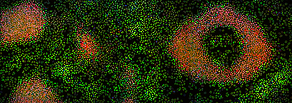
When cells are created, they have the potential to take on many different types of functions. Jean Cook’s lab studies the process of how a cell determines what its future function should be, and how it transforms itself to fit that function. In An Eye on the Future (below), Cook shows the process at work: The green cells have committed to becoming muscles, while the red cells are still deciding what they want to be when they grow up. The image looks like abstract art, and it may help Cook’s team develop methods for repairing damaged tissue or halting the growth of cancerous cells.